
 Deutsch
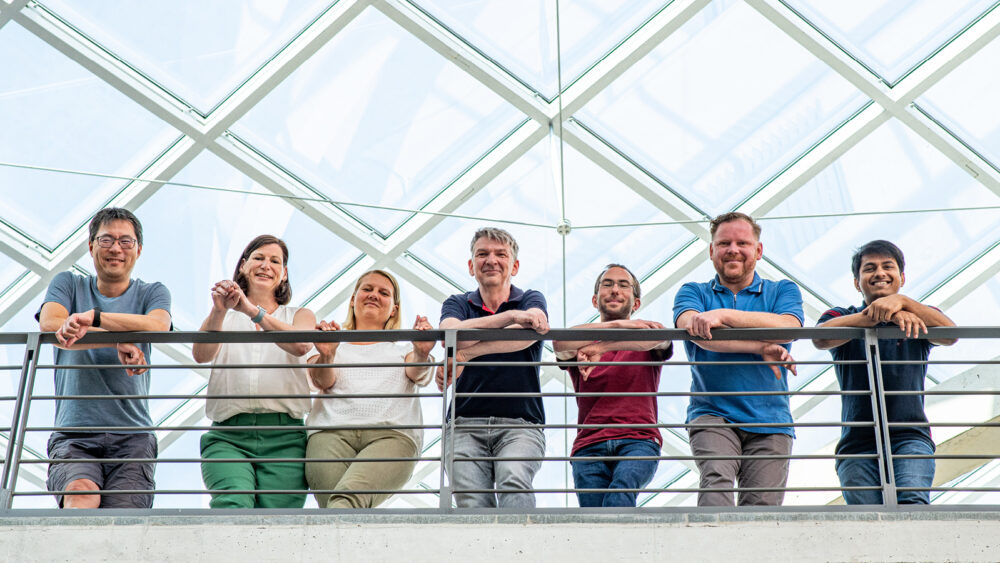
Flow Cytometry & Cell Sorting (FCCF)
State-of-the-art cell analysis and cell sorting through technical innovation
The Flow Cytometry Core Facility (FCCF) was established in 2000 as a Shared Resource Lab of the DRFZ, the Charité – Universitätsmedizin Berlin, and the Max Planck Institute for Infection Biology. With a comprehensive range of services and proprietary technological innovations, the FCCF offers state-of-the-art analyses and sorting of cells.
Methods and applications
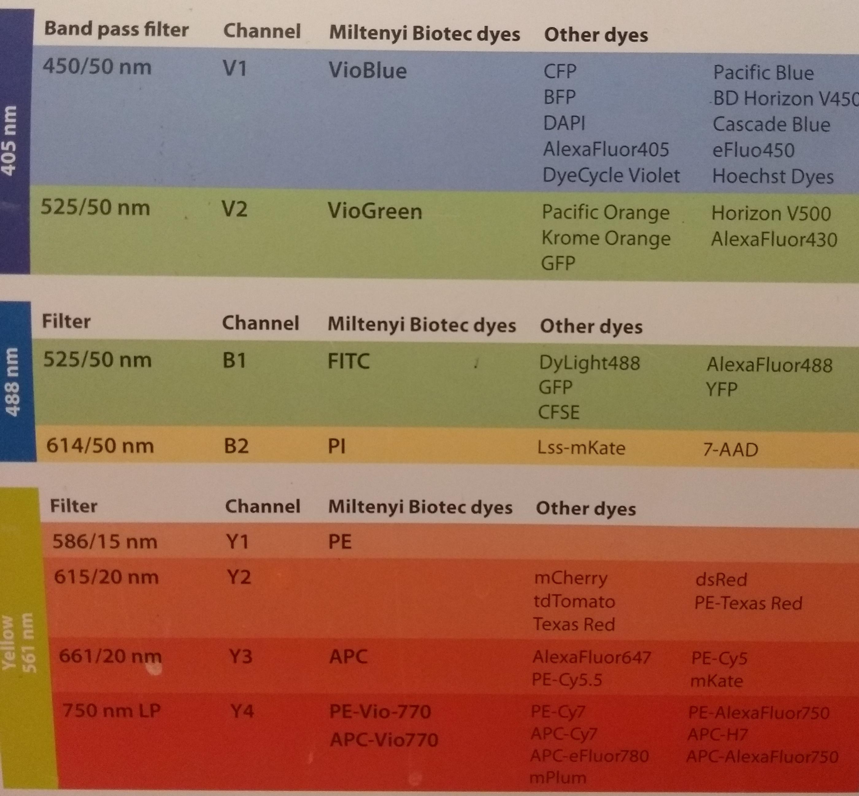
Flow cytometry is an analytical method for the quantitative recording of physical, biochemical/cell biological and immunogenetic parameters of individual cells. On this basis, various cell features can be determined. The flow cytometers of the FCCF allow the simultaneous measurement of up to 28 characteristics of a cell at a throughput of up to 20,000 cells per second. In such a “multicolour” approach, it is possible to e.g. analyse cells of patients before and during therapy in order to monitor the success of therapy, but also to investigate the cellular processes underlying rheumatic inflammation.
In addition to the cytometric analysis of cell types of interest, cells can also be isolated for further molecular, genetic and biochemical analyses. For this purpose, cells are first analysed (as in the cell analyser) and then packed into individual droplets. These are given a specific electrical charge and are then directed into collection tubes within an electrical field. With specially adapted sorting protocols, even very sensitive cell types, such as stroma or plasma cells, or very rare cell types, such as autoreactive lymphocytes (in combination with pre-enrichment methods, such as magnetic cell sorting), can be analysed or sorted.
Basic flow cytometry course
Once a month, we offer a two hour basic flow cytometry course on the proper use of the techniques used in flow cytometry. The course is open to all who are interested. The dates will be announced or can be requested by email.
Service and cytometry introduction course
All new users must fill out the registration form and attend the cytometry introduction course. The course takes place monthly. Please contact the staff for detailed information.
Technological developments and innovations
To increase the number of parameters of cells that can be detected by flow cytometry, we are investigating cytometric pulse shape analysis in collaboration with the Berlin-based company APE. In this method, the resulting pulse shape of the scattered light from a cell is detected in an angle-dependent manner and classified by data processing. This allows the discrimination of cell properties based on the pulse shape alone, i.e. it is not necessary to label the cells with a surface marker, for example. In a further step, this method will be extended to enable cell sorting.
Also in cooperation with the Berlin-based company APE, we have developed the LED-based calibration tool for flow cytometers (quantiFlashTM). It enables us to quantify the sensitivity of flow cytometers.
We introduced an experimental strategy, in which discontinuous emission spectra were acquired and analyzed by principal component analysis (PCA). In a proof-of-principle study we could show, that the simultaneous detection of up to 4 fluorochromes, which were all excited by a 488 nm Argon-laser could be resolved by the detection of a discontinuous emission spectrum. In other words we use a standard cytometer just by changing the optical filters as a multispectral cytometer. No further expensive modifications as described in the literature are required.
We have developed a flux reactor that sterilizes the sheath fluid at the point of use. The flux reactor consists of an array of 6 UV-C LEDs. Such UV-C radiation source is also used in water treatment and disinfection. The module is currently installed in an INFLUX cell sorter and allows to reduce the number of microorganisms by up to 1000 times. The module was manufactured in cooperation with APE.

We developed a cooling device in order to keep the sheath fluid temperature on a FACS Aria constant. We found that the foccusing of the side-streams on a FACS Aria are temperature sensitve. Room and instrument temperature shifts of more than +/- 5K results in a defocusing of the side-streams and the recovery of sorted cells decreased drastically. Furthermore, the sensitivity of delayed lasers decreased due to instable laser-delay.
To overcome this effects we hold the sheath-fluid constant at 17°C by using our device.

During long-term sorts cells often sediment at the bottom of the sample tube. In consequence, the threshold rate are instable or interupted and cells in the settlement are exposed to cytotoxin. In cooperation with Advalytix we modified their universal acoustic mixing platform “SAWStation4” so that we can implement it in a FACS Aria.
In our study we could show that the yield of sorted cells was about 5% higher compare to cells mixed by the original FACS Aria agitator. Moreover, we found less dead cells in sorted populations.

Among others, the group is supported by the State Berlin and the European Commission (research project PULZY).
![]()
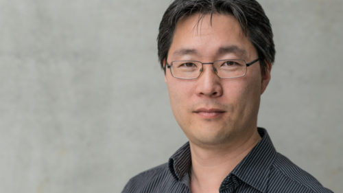
Group leader
Prof. Dr. Hyun-Dong Chang
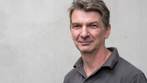
Technical Manager
Dipl. Ing. Toralf Kaiser
Operators
Dipl. Biochem. Jenny Kirsch
Dr. Kerstin Heinrich
Scientists/project team members
Dr. Daniel Kage
Dr. Alexander Wolf
• Max Planck Institute for Infection Biology, Berlin
• Technische Universität Berlin
• Angewandte Physik und Elektronik GmbH (APE), Berlin
• MPI für molekulare Genetik, Berlin
• Physikalisch-Technische Bundesanstalt, Berlin
Patente/Patents
- Kaiser, J. Kirsch,
A. Grützkau,
Patent Number: 9958393
‘Principle component analysis (PCA) – based analysis of discontinuous emission spectra in multi-chromatic flow cytometry’ - Kaiser, T. Kolbe, M. Kneissl,
Patent Number: 9498550
‘Flow cytometer desinfection module’ - Kristen Feher, Toralf Kaiser, Konrad von Volkmann, Sebastian Wolf
Patent Number: US10337975B2
‘Method and system for characterizing particles using a flow cytometer’
Ausgewählte Publikationen/Selected Publications
Giesecke, K. Feher, K.v. Volkmann, J. Kirsch, A. Radbruch, T. Kaiser, “Determination of background, signal-to-noise and dynamic range of a flow cytometer – a novel practical method for instrument characterization and standardization”, Cytometry Part A 91.11 (2017): 1104-1114.
Feher, K. von Volkmann, J. Kirsch, A. Radbruch, J. Popien, and T. Kaiser, “Multispectral flow cytometry: The consequences of increased light collection,” Cytometry A, Jun. 2016.
Comparison study of various flow cytometers using a novel ultra stable calibration light source. von Volkmann, K., Feher, K., Popien, J., Kaiser, T. Poster at DGfZ meeting, Berlin, 2015.
Classification of Flow Cytometry Samples with a Dimension Reduction and Binning Approach. Feher, K., Radbruch, A., Kaiser, T. Poster at Cyto conference, Glasgow, UK, 2015
- 1 Analyser MACSQuant (3-lasers) bobby_configuration
- 1 Analyser MACSQuant (3-lasers) MACS Quant Camilla
- 1 Analyser LSRFortessa (4-lasers)Wayne 6v2b4y3r 17022016
- 1 Analyser FACS Canto (3-lasers) Canto New
- 1 Analyser Symphony A5 (5-lasers) Janice 5b8v3r5yg7uv 23022017 1
- 2 Cell Sorter FACS Aria II (4-lasers) AriaII Floyd/Robin
- 1 Cell Sorter Influx (5-lasers)
- 1 Cell Sorter Sony MA900 (4-lasers) Konfiguration Sony MA900